
Updated: 17 November 2026 la 19:54. Articolul a fost revizuit pentru a reflecta informațiile disponibile la această dată.
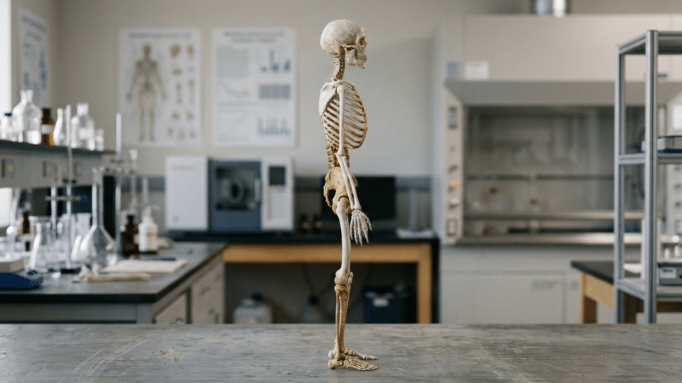
The shape of the human skeleton cannot be reduced to a single mutation or a hypothetical “bipedal-walking gene”. A study published in Nature on 23 September 2026 found that differences between humans and other great apes include thousands of changes in DNA sequences that regulate the activity of genes involved in cartilage and bone formation.
Related: Why do large dogs age faster? A genetic study of 894 dogs explains the role of “jumping genes”, with further relevant information.
The key finding is not that researchers identified a single genetic instruction for the pelvis, spine or knee. They produced an experimental map of many small changes distributed across the genome that can alter when, where and how much a gene is activated in cells involved in skeletal development.
In brief: the evolution of the human skeleton appears to have depended largely on gene regulation, not only on changes in protein-coding genes. The study also offers a relevant clue about articular cartilage, but it does not establish the cause of osteoarthritis and currently has no direct medical application.
Human skeletal evolution is also written in genetic “switches”
A gene does not operate continuously, or at the same intensity, in every cell. It is surrounded by regulatory sequences, including promoters and enhancers, that influence its activation. As a useful approximation, they can be compared to a control system: they do not necessarily change the part produced by a factory, but can alter the timing, location and volume of production.
This distinction matters in the evolution of the body. Two species may have very similar genes for proteins involved in bone and cartilage, yet use those genes differently during development. A genetic signal activated slightly earlier or later, or in a different cell population, can influence the tissues from which bone and joint structures develop.
The authors of the Nature paper tested 561,410 substitutions that arose along the human evolutionary lineage in promoters and enhancers. Among the regions analysed, 15,077 loci showed human-specific regulatory activity in the experimental systems used.
This figure should not be read as the number of “skeleton genes”. A locus is a position or region in the genome, and many such regions may act on the same gene or on genes in the same biological pathway. The underlying idea is polygenic: numerous regulatory adjustments, each with a potentially modest effect, can over time produce substantial anatomical differences.
What the researchers tested, and why the method matters
The first part of the study used a test known as an MPRA, short for massively parallel reporter assay. In practical terms, the method makes it possible to test a very large number of DNA fragments simultaneously: researchers can see whether a human version of a sequence affects the activity of a molecular signal more or less than the version being compared.
MPRA is well suited to the question: “Does this sequence have different regulatory activity in these cells?” It cannot on its own answer the broader question: “Did this difference change the shape of a bone in a living organism?” Biological steps between those two claims must be demonstrated separately.
The authors supplemented the assay with human-chimpanzee and human-gorilla hybrid cells, differentiated into osteochondral progenitors—precursor cells for bone and cartilage. The advantage of this approach is that alleles from the two species can be studied in the same cellular environment. This allows researchers to better separate the effect of the DNA sequence itself, known as a cis-regulatory effect, from effects produced by the rest of the cellular environment.
It is a strong experimental control, but it is not a skeleton growing in a body. Cultured cells do not reproduce all the mechanical forces, hormonal signals, tissue interactions and developmental stages that shape a real joint. This is one limitation that must remain visible when interpreting the findings.
Cartilage is not simply a “cushion”: the role of glycosaminoglycans
The study’s most concrete biological observation concerns the extracellular matrix, the material surrounding cells that gives tissues their structure and mechanical properties. In cartilage, this matrix is particularly important: it must withstand compression while allowing joints to move.
The paper reports remodelling in pathways involved in the extracellular matrix and in the biosynthesis of glycosaminoglycans, abbreviated as GAGs. Glycosaminoglycans are chains of complex sugars generally attached to proteins in the matrix. They help cartilage retain water and withstand pressure.
In the joint tissues analysed, the authors reported GAG content approximately three to four times lower in humans than in non-human great apes. This is a difference between species, not an analysis of individual disease risk in humans.
| Claim | What the data support | What does not automatically follow |
|---|---|---|
| Many DNA regions have different regulatory activity | The study identified 15,077 loci with human-specific activity in the systems tested. | That every region produces a visible anatomical trait. |
| Cartilage-matrix pathways are remodelled | The findings indicate regulatory changes related to the extracellular matrix and GAGs. | That humans have “defective cartilage” or an inevitable disease. |
| Humans had fewer GAGs in the samples compared | The authors report a roughly three- to fourfold difference from other great apes. | That this difference causes osteoarthritis. |
The connection with joint disease warrants explanation precisely because it is tempting. Studies of human cartilage affected by osteoarthritis show that changes in, and loss of, glycosaminoglycans are relevant to the degradation of the cartilage matrix. For example, a mechanistic study of GAG synthesis in osteoarthritis described the loss of glycosaminoglycan chains as a phenomenon associated with the pathological process.
But a resemblance between two phenomena does not automatically establish a single cause. A different GAG level between species that emerged through evolution is not equivalent to the degradation process that occurs in a diseased joint over a lifetime. The direction of causality, the precise mechanical effect and the contribution of each genetic variant remain to be tested.
Why the study does not, by itself, explain osteoarthritis
Osteoarthritis is a disease of the whole joint, not cartilage alone. According to the World Health Organization, associated factors include age, joint injuries and overuse, excess weight, genetics and certain metabolic diseases.
The work on skeletal evolution therefore opens a biological hypothesis: certain evolutionary features of the joint matrix may deserve investigation as part of human susceptibility to joint degeneration. It does not, however, provide a risk test for an individual, a new prevention method or a validated therapeutic target.
There is also an independent line of research that makes caution more useful than sweeping conclusions. An earlier study published in Cell, available through PubMed Central, analysed regulatory elements in knee chondrocytes. Its authors found signals of evolutionary selection and constraint in these regions, as well as an enrichment of variants associated with osteoarthritis risk.
That study included functional validation of an enhancer at the GDF5 locus, including in mouse models. It supports the broader idea that genetic regulation of joint development may be relevant to risk. However, it did not test the atlas of loci from the Nature study or the mechanism involving GAGs. This is conceptual convergence, not a direct replication.
What this map changes, and what remains to be learned
The new research shifts attention away from the search for a single genetic explanation and towards an architecture made up of many regulatory changes. In this framework, the evolution of the human skeleton does not mean that a protein was rewritten from the ground up, but that existing networks were adjusted in different cells and at different moments in development.
Another paper, published in 2026 in Genome Biology and Evolution, likewise reported differences in activity distributed across thousands of regulatory elements involved in postcranial skeletal development, comparing human and chimpanzee sequences. Its findings, summarised in the study’s bibliographic record, support the general model of polygenic evolution. The methods and regions analysed differ, however, so the figures should not be combined as though they described exactly the same experiment.
To link a DNA change more firmly to an anatomical trait would require, among other things, experiments that selectively alter individual variants, developmental models closer to intact tissue, and measurements of effects on structure and biomechanics. Establishing a connection with disease would then require clinical and population data, rather than comparisons between species or cultured cells alone.
Editorial method and the finding’s practical limit
This InfoBuz article synthesises the primary Nature paper, an independent study of knee regulation, separate research on regulatory elements in postcranial development, and the clinical context provided by the WHO. The figures for 561,410 substitutions, 15,077 loci and the GAG difference come exclusively from the Nature study, not from press releases or secondary reporting.
The research is valuable for its explanatory power: it provides a better map for the question of how the human skeleton formed. It does not change the diagnosis or treatment of joint diseases today. For readers, the most accurate conclusion is that DNA regulating genes can be as important as genes themselves when trying to understand why the human body has its particular form.
Frequently asked questions
Has the gene for bipedal walking been discovered?
No. The study points to many changes in sequences that regulate genes active in cartilage and bone development. It does not identify one gene responsible for bipedal walking.
What are glycosaminoglycans?
They are chains of complex sugars in the extracellular matrix. In cartilage, they help tissue retain water and withstand compression in joints.
Does the study show that humans develop osteoarthritis because of evolution?
No. The finding offers a biological clue worth investigating, but osteoarthritis is multifactorial and depends, among other things, on age, injuries, weight, mechanical stress, genetics and metabolic diseases.
Why did the researchers use hybrid cells from different species?
Hybrid cells allow human and other great ape alleles to be compared in a shared cellular environment. This means that observed differences can be attributed more clearly to the DNA sequence, rather than only to the cellular environment.
Sources consulted
- The gene-regulatory evolution of the human skeleton
- In Vitro Massively Parallel Screening of Human Regulatory Elements Involved in Postcranial Skeletal Development for Differential Activity Compared to Chimpanzee
- Evolutionary Selection and Constraint on Human Knee Chondrocyte Regulation Impacts Osteoarthritis Risk
- Xylosyltransferase-I regulates glycosaminoglycan synthesis during the pathogenic process of human osteoarthritis
- Osteoarthritis


Discuția cititorilor
Ai o opinie sau o întrebare despre acest articol? Spune-ne ce crezi.
Fii primul care deschide discuția.
Te rugăm să păstrezi un limbaj civilizat și să discuți la subiect. Comentariile spam sunt filtrate automat.