
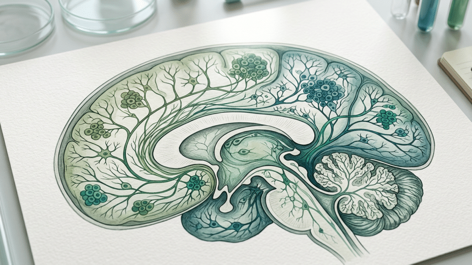
A study published in Nature found cortical neurons with extensive chromosomal changes more often in prenatal samples than in samples collected after birth. The authors propose that some of these cells are selected and eliminated around the time of birth, as brain circuits form and are refined.
The headline captures the broad idea, but the precise wording matters: the researchers did not follow the same cell before and after birth. They compared post-mortem samples from different ages. The data indicate a perinatal decline in the frequency of some neurons with extensive genetic changes; cell elimination is the biological interpretation supported by several types of analysis, not a direct recording of the process.
In brief: this is fundamental research into brain development. It is not a pregnancy test, cannot estimate an individual child’s risk, and does not show that such changes explain autism, epilepsy, schizophrenia or other diagnoses.
What researchers measured in neurons with CNVs
The study, available in the Nature research article, analysed the genomes of 2,357 individual neurons from the human cortex, obtained from fetal, childhood, adult and older-adult post-mortem tissue. The team used two single-cell genome amplification and sequencing techniques.
The main focus was CNVs, short for copy-number variants: variations in the number of copies of segments of DNA. Rather than being present in the usual number of copies, a genetic section may be duplicated or lost in a cell. When the change arises after fertilisation, it may be carried by only some of a person’s cells; this is known as genetic mosaicism.
Mosaicism does not automatically mean disease. The National Human Genome Research Institute explains that people with mosaicism have cells with different genetic profiles, and the consequences can vary widely or be absent. A change detected in one neuron is not equivalent to an abnormality present in all of a fetus’s cells, nor is it a prenatal test result.
The difference observed between prenatal and postnatal samples
Under the authors’ technical definition, 25.9% of the prenatal cortical neurons analysed had extensive CNVs affecting regions on multiple chromosomes. The reported average proportion in samples collected after birth was about 6.2%.
The difference matters, but it should not be read as a measurement of “a quarter of every fetus’s brain”. It is an average derived from a sample of individual cortical neurons taken from post-mortem specimens, using specific rules for CNV classification. The prenatal range reported by the authors, from 12.7% to 46%, also shows the variation between samples.
The main result pointed in the same direction with both sequencing methods used in the study. This provides a useful internal technical check. It is not, however, the same as replication of the conclusion by an independent laboratory using different samples and technologies.
Why “around birth” is more accurate than “before birth”
The authors do not argue that every neuron with an altered genome disappears before birth. They found that neurons with extensive CNVs were far more common in fetal than in postnatal samples, and link that difference to the known period of intensive brain remodelling.
In a multi-omic analysis of 44,861 nuclei, neurons with a high CNV burden showed disruption in gene modules associated with synapses. A postnatal subgroup linked reduced expression of certain synaptic genes with greater activation of pathways related to cell death. The researchers also reported micronuclei and aberrant genetic material in prenatal samples, with this signal declining with age.
These findings converge on the hypothesis of selection: cells with greater genomic imbalances may be at a disadvantage when attempting to integrate into brain networks. But an association does not establish direct causation. The study does not prove that synaptic disruption causes the death of a particular cell, and it does not follow that cell over time.
What earlier studies confirm — and where differences remain
The existence of somatic variation in neurons is not a new idea. Earlier studies found mosaic CNVs in human neurons, including in the frontal cortex. For example, research published in 2013 reported megabase-scale CNVs in 13%–41% of the neurons analysed, depending on the sample and method.
Other findings have been more cautious. A 2014 study published in Cell Reports estimated that at least 95% of neurons in the normal brain tissue analysed were euploid, meaning they did not show major changes in chromosome number. The findings do not necessarily conflict: the studies used different samples, detection thresholds, brain regions and definitions of CNVs.
For more information on medical research, see also Graphene and stroke: experimental sensors identified vulnerable brain tissue in real time in a mouse study, which offers complementary details on this subject.
This is one of the field’s decisive limitations. Single-cell sequencing requires the DNA available in one cell to be amplified before analysis. The amplification method, sequencing coverage, reference genome and calling algorithm can alter the estimated CNV rate. A methodological study published in 2023 showed precisely that the choice of technique can change the profile of detected variants.
What the study does not say about pregnancy and neurodevelopment
The finding does not provide a medical tool for pregnant people. Based on this research, there is no clinical test that measures cortical neurons with CNVs during pregnancy, nor is there a recommended intervention intended to influence the proposed cell selection.
It is also not appropriate to translate the finding directly into a diagnosis of autism, epilepsy or intellectual disability. Some CNVs can have clinical relevance in certain contexts. There is, for example, a Romanian cohort of patients assessed for global developmental delay and/or intellectual disability in which pathogenic CNVs were investigated. But those clinical data concern children selected for medical assessment and do not test the phenomenon observed in individual neurons in the Nature study.
The distinction matters: a pathogenic CNV identified through clinical genetic testing may be present in many cells throughout the body. A somatic CNV detected in a single neuron in brain tissue is a cell-biology observation that may not be present in blood, saliva or the rest of the body.
What the next steps in research should show
Robust confirmation would require independent samples analysed by other laboratories, across more populations and cortical regions. More varied gestational stages, the effects of the post-mortem interval and sample composition, and independent methods of CNV detection would also need to be tested separately.
The study adds a coherent hypothesis to existing knowledge about brain development: the formation of neuronal connections may also act as a filter for certain genetic imbalances that arise in cells. For now, the most cautious conclusion is that the data support incomplete, probably predominantly perinatal selection — not a total “clean-up” of the brain, and not a ready-to-use clinical explanation.
Frequently asked questions
What are CNVs?
CNVs are variations in the number of copies of DNA segments. A genetic region may be duplicated or lost in some cells. When they arise after fertilisation, they can produce genetic mosaicism.
Does the study mean that a quarter of the fetal brain is abnormal?
No. The 25.9% figure is an average for individual cortical neurons in a set of post-mortem samples, defined according to the study’s technical criteria. It does not measure the proportion of the entire brain in an individual fetus and is not a diagnosis.
Did the study prove that abnormal neurons die before birth?
Not directly. The study compares prenatal and postnatal samples and finds a lower frequency after birth. The authors support the hypothesis of predominantly perinatal elimination with associated molecular analyses, but they did not track the same cell over time.
Is there a prenatal test based on this discovery?
No. The study is fundamental research using post-mortem brain tissue and does not provide a screening test, diagnostic test or treatment for pregnancy.
Does the finding explain autism or other neurodevelopmental disorders?
No. The research may inform future questions about brain development, but it does not show that the observed mechanism causes autism, epilepsy, schizophrenia or intellectual disability.
Sources consulted
- Nature — Perinatal elimination of genetically aberrant human cortical neurons
- PubMed — Mosaic copy number variation in human neurons
- PubMed — Single-Cell, Genome-wide Sequencing Identifies Clonal Somatic Copy-Number Variation in the Human Brain
- PubMed — Single-cell somatic copy number variants in brain using different amplification methods and reference genomes
- National Human Genome Research Institute — Mosaicism




Discuția cititorilor
Ai o opinie sau o întrebare despre acest articol? Spune-ne ce crezi.
Fii primul care deschide discuția.
Te rugăm să păstrezi un limbaj civilizat și să discuți la subiect. Comentariile spam sunt filtrate automat.